 While many conditions may cause memory loss, Alzheimer's disease is the predominant cause, and early and accurate diagnosis enables the most efficacious treatment. Early identification also allows families and patients to plan for the future while the patient can still be involved in the decision making process. While many conditions may cause memory loss, Alzheimer's disease is the predominant cause, and early and accurate diagnosis enables the most efficacious treatment. Early identification also allows families and patients to plan for the future while the patient can still be involved in the decision making process.
Despite the common assertion that autopsy is the only method of certain AD diagnosis, it should be noted that the NINCDS-ADRDA diagnostic criteria for Alzheimer's disease are more than 90% accurate, even at early stages of the disease. This diagnostic process includes:
Medical history should query for initial symptoms that characterize specific dementing disorders. Examples include family history of dementia, risk factors for Alzheimer's disease, visual hallucinations, loss of balance and marked fluctuation in level of alertness or confusion, marked reduction in speech or marked change in personality or behavior, and short-term memory loss.
Laboratory tests should be done to diagnose and treat other possible causes of mild cognitive impairment or dementia such as uncontrolled diabetes, vitamin B12 deficiency and thyroid disease.
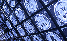
Neuroimaging should be done to identify hemorrhage, stroke, hydrocephalus, tumor, and degenerative disorders with selective patterns of atrophy. In Alzheimer's disease, the entorhinal cortex and hippocampus in the medial aspect of the temporal lobe are the first cortical areas to show neuronal pathology and cell loss. Therefore, hippocampal atrophy that is greater than that of the remaining cortical areas is virtually diagnostic of Alzheimer's disease, particularly if the atrophy is not explained focal ischemia, hydrocephalus, a long history of poorly controlled seizures, tumor or focal trauma.
If the patient is in the mild cognitive impairment (MCI) stage then an MRI is needed to detect subtle amounts of atrophy. If a patient has very early stage impairment, a PET scan in addition to an MRI may be required. Decreased tissue activity, identified by PET scan, precedes structural abnormalities, identified by an MRI, by two or more years. However, if the patient is demented, then the amount of atrophy is usually substantial and a CT scan of the brain is adequate to detect selective atrophy, particularly if a spiral CT scan is done with coronal cuts.
|